The kidneys are a pair of bean-shaped organs found in vertebrates and some invertebrates that function to maintain water balance and also excrete metabolic wastes. The human kidney is located below the diaphragm and behind the peritoneum. It is usually 10-12 cm long and is made up of repetitive units called nephrons.
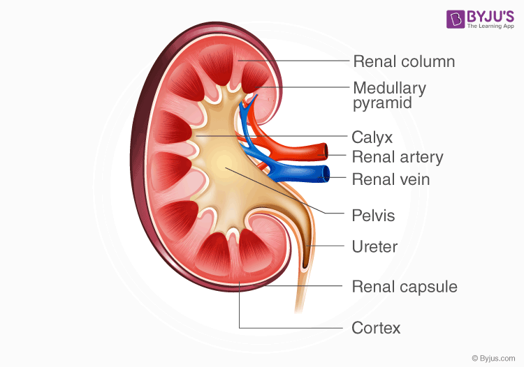
Find out the simple diagram of the human kidney below with all the parts explained.
Well-Labelled Human Kidney Diagram

Structure of the Kidney
- The bean-shaped structure of the kidney is composed of both concave and convex borders.
- A small curvature called renal hilum is present on the concave face of the kidney from where the renal artery enters the kidney, and renal vein and ureter leave the kidney.
- The renal capsule is a tough fibrous tissue that surrounds the kidney and is itself covered by a layer of perirenal fat. This capsule protects the kidney from outside trauma and injury.
- The kidney is divided into two major zones – the outer renal cortex and inner renal medulla.
- The renal cortex surrounds a portion of the medulla to form the renal pyramids.
- Pelvis is a funnel-shaped part of the ureter that extends from the kidney and is formed by the union of major and minor calyces (sing: calyx).
- The renal calyces are present surrounding the renal pyramids. Calyces are chambers through which urine passes.
- The renal artery supplies blood to the kidney, whereas renal veins drain the kidney blood and further connect it to inferior vena cava.
- The nephron is the functional unit of the kidney that traverses through both the medullary and cortex regions.
- The nephron filters, reabsorbs, secretes and excretes the supplied blood to convert it into urine.
- The nephrons are composed of the following parts – glomerulus, Bowman’s capsule, distal convoluted tubule, proximal convoluted tubule and collecting duct.
- The kidneys are important for the process of urine formation and excretion of other metabolites and form a major part of the urinary tract.
Nephrons – Diagram and Structure
Nephron is a microscopic structure that is the functional unit of the kidney. There are about 1,000,000 nephrons in each human kidney. Let us look at the components of the nephron. Below is a well-labelled and easy diagram of the nephron for your better understanding.

- Broadly, a nephron can be divided into two parts – renal corpuscle and renal tubule. The renal corpuscle is the filtering component and renal tubules carry the filtered liquid away.
- A human nephron is a long fine tubule that is about 30-55 mm long.
- One end of this long tubule is closed and folded into a double-walled cup-like structure called the Bowman’s capsule.
- Inside the capsule are present a number of fine blood capillaries that are known as glomerulus. Both the capsule and glomerulus constitute the renal corpuscle.
- The glomerulus receives the blood supply from afferent arterioles. Here water and solutes are filtered out of the blood and transported to Bowman’s capsule.
- Fluids from the blood, ultra filtered in the several layers of the glomerulus, (and now called filtrate) are finally passed onto the nephron tubule via Bowman’s capsule.
- The nephron tubule is made up of a proximal convoluted tubule, loop of Henle, distal convoluted tubule and the collecting duct.
- The filtrate passes through the tubule where certain substances are secreted into the fluid and water is selectively reabsorbed from it to form urine.
Stay tuned to BYJU’S Biology for more updates.
Also Read:
Comments