Find below the important notes for the chapter, Locomotion and Movement, as per NEET Biology syllabus. This is helpful for aspirants of NEET and other exams during last-minute revision. Important notes for NEET Biology- Locomotion and Movement covers all the important topics and concepts useful for the exam. Check BYJU’S for the full set of important notes and study material for NEET Biology and solve the NEET Biology MCQs to check your understanding of the subject.
Download Complete Chapter Notes of Locomotion and Movement
Download Now
| Name of the NEET sub-section | Topic | Notes helpful for |
| Biology | Locomotion and Movement | NEET exams |
Recommended Videos:


Don’t miss: Answer Key NEET 2022
Locomotion and Movement – Important Points, Summary, Revision, Highlights
Locomotion and Movement
Movement is a characteristic of all the living organisms. From the protoplasmic movement in a cell and unicellular organisms to the movement of organs in multicellular organisms, movement accounts for the various important functions in the living organisms.
The movement, which leads to a change in the place is called locomotion.
There are three types of movement in a cell and organs. These are:
- Amoeboid movement- It is like that of pseudopodia in amoeba, e.g. macrophages, leukocytes and even cytoskeletal microfilaments show ameboid movement.
- Ciliary and Flagellar movement- Ciliary movement in the epithelial lining of the trachea, reproductive tract, etc. Sperms show flagellar movement.
- Muscular movement- Muscles are responsible for most of the movement in a multicellular organism. Breathing, the function of heart, digestion, movement of appendages, locomotion, everything is performed by various muscles in our body. Locomotion is the coordinated movement of skeletal, neural and muscular systems.
Muscles originate from the mesodermal germinal layer. Muscular tissues have certain unique properties such as contraction, extension, excitation, elasticity, etc.
We all know, there are three types of muscles:
Cardiac muscles- striated and involuntary, present in the heart.
Visceral muscles- smooth and nonstriated. They are also involuntary and support various internal organs and take part in various functions such as digestion, reproduction, etc.
Skeletal muscles- striated and voluntary, responsible for the locomotion and movement of appendages.
Let’s learn in more detail about skeletal muscles, their structure and mechanism of muscular contraction.
Anatomy of Muscle Fibre and Sarcomere
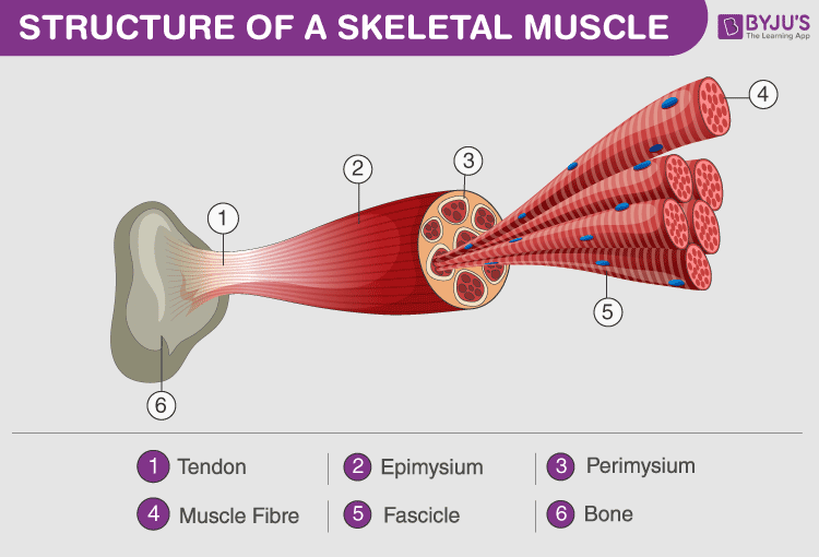
Skeletal muscles are the most abundant muscles. They are made up of bundles of muscle fibres wrapped around by connective tissue.
Fascicles (Muscle bundle)
A muscle such as a biceps comprises many muscle bundles (fascicles) held together by fascia a connective tissue layer. Each fascicle contains many muscle fibres.
Muscle Fibres
Muscle fibres are elongated cells. They are organized into bundles called fascicles. The characteristics features of muscle fibres are:
- Each skeletal muscle fibre is long, cylindrical and striated.
- It is syncytium, i.e. contains many nuclei.
- Sarcolemma- is the plasma membrane of the muscle fibre.
- Sarcoplasm- is the cytoplasm of the muscle fibre.
- Sarcoplasmic reticulum- is the endoplasmic reticulum of the muscle fibre. The storehouse of Ca ions.
Myofibrils
Each muscle fibre has multiple myofibrils running lengthwise and parallel to each other in the sarcoplasm. The alternating dark and light bands present in myofibrils give the muscle striated appearance. Each myofibril consists of even simpler structures called myofilaments.
Myofilaments
There are two types of myofilaments present in a myofibril. Thin and thick filaments. Thin filaments and thick filaments have to attach to each other for muscle contraction.
a. Thin filament or Actin filaments are made up of three different proteins.
- It contains two polymeric filamentous or ‘F’ actin filaments, which are wound together. They are a polymer of globular or G actin monomers.
- Tropomyosin filaments are coiled around the actin filaments lengthwise.
- Troponin, which is present at certain points on the tropomyosin, covers the active binding sites for myosin.
- Troponin and tropomyosin regulate the binding of actin and myosin filaments, hence play an important role in muscle contraction.

b. Thick filament or Myosin filaments are made up of myosin.
- It is a polymeric protein consisting of monomeric units of a protein known as meromyosin.
- It has three parts, tail, short arm or neck and a globular head.
- The head and cross arm protrude outwards from the filament at regular intervals.
- The globular head contains binding sites for ATP and actin.
- The head acts as an ATPase enzyme.
Sarcomere, the functional unit of muscle contraction
A myofibril contains hundreds of sarcomere, which are connected end to end.
A sarcomere is the basic unit of muscle contraction. It contains repeating units of actin and myosin filaments in an orderly fashion.
- Sarcomeres are joined at the end by interweaving filaments forming the ‘Z’ line. It is the limiting membrane of a sarcomere.
- Actin filaments or thin filaments are attached to the Z line at frequent intervals.
- Thick filaments or myosin filaments are present between the thin filaments. They are held together at their place by a very thin fibrous membrane known as ‘M’ line.
- The actin and myosin filaments overlap each other in a specific pattern and result in the striation pattern of muscles. They form 3 types of bands.
- ‘I’ band or Isotropic band- This is the light band and comprises actin filaments attached to the two adjacent sarcomeres. Here, thick and thin filaments do not overlap.
- ‘A’ band or anisotropic band- This is the dark band, it contains overlapping actin and myosin filaments.
- ‘H’ zone is the middle portion of the thick filaments, where thin filaments do not overlap the thick filaments.
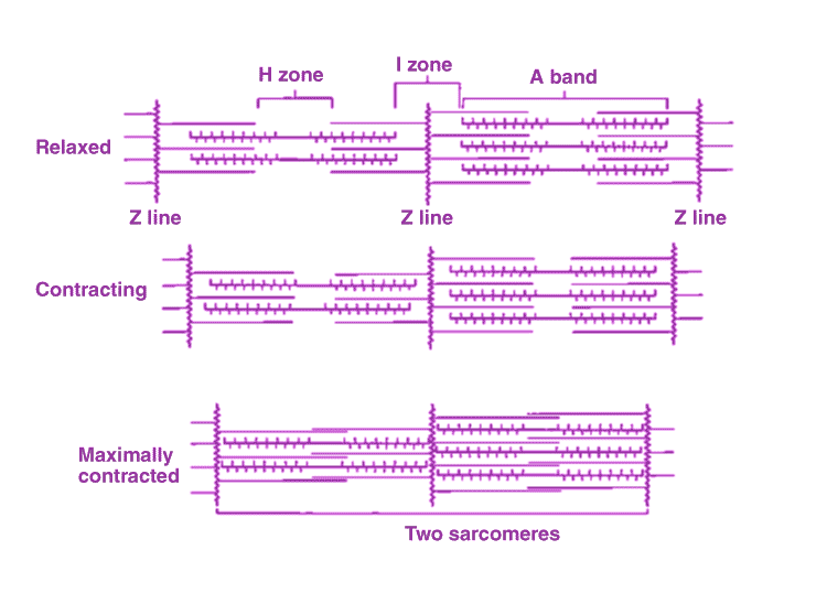
To summarize, the sarcomere is the basic unit of muscle, made up of thick and thin myofilaments. Hundreds of sarcomere are present in myofibril. Multiple myofibrils form a muscle fibre. A bundle of muscle fibres is known as fascicles. Multiple muscle bundles are enclosed in a fascia and constitute a muscle.
Let us now understand the complex physiology of muscle contraction.
Mechanism of Muscle Contraction
Muscle contraction is the result of the sliding of actin and myosin filaments over each other.
Sliding filament model was proposed by Andrew and Hugh Huxley.
Sliding causes an increase in the overlap of thick and thin filaments and shortening of the sarcomere. This results in the contraction of muscles.
Muscle Contraction Steps
- Brain or spinal cord (CNS) transmits a signal through motor neurons for the initiation of muscle contraction.
- The neural signal causes the release of the neurotransmitter, Acetylcholine at the synaptic cleft of the neuromuscular junction. Acetylcholine binds to receptors present on the muscle fibre and causes depolarization of sarcolemma.
- The action potential thus generated spreads through the muscle fibre. Ca2+ ions are released in the sarcoplasm from the sarcoplasmic reticulum. The release of Ca2+ is regulated by another protein dystrophin. The dystrophin coding gene is the longest gene in the human body.
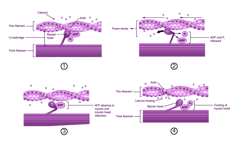
- The Ca2+ ions bind to troponin and cause conformational changes. The myosin-binding sites present on actin filaments become exposed.
- Myosin head also contains a binding site for ATP, where ATP binds. ATP hydrolysis takes place by ATPase activity of the myosin head. The energised myosin head (cocked) binds to the active binding sites on actin forming a cross bridge.
- The attachment is followed by the release of phosphate from the myosin head triggering the ‘power stroke’. Myosin filaments bend and pull actin filaments towards the centre of the sarcomere causing shortening of the sarcomere and muscle. ADP is released in the process.
- The detachment of myosin head is also driven by ATP.
- In the presence of sufficient Ca2+ ions, the process repeats again and again.
Muscle Relaxation
Once the neural signal ceases. Acetylcholinesterase inactivates acetylcholine in the synaptic cleft. Muscle fibres come to a resting state. Ca2+ ions are pumped back into the sarcoplasmic reticulum. In the absence of Ca2+ ions, the troponin-tropomyosin complex again covers the myosin-binding sites on actin filaments. Z line of sarcomere returns to its original position and muscles become relaxed.
Muscle Fatigue
Muscle contraction is powered by ATP. Muscle fibres get ATP from the storage of creatine phosphate and glycogen. Under normal conditions, the glycogen is converted to glucose, which further releases ATP in cellular respiration.
In the case of strenuous exercise, muscles require high amounts of energy. The body could not supplement the oxygen demand and glucose is broken down anaerobically. This results in the accumulation of lactic acid causing muscle fatigue.
Rigour Mortis, the temporary stiffening of muscles after death is caused due to depletion of ATP as the cellular respiration ceases. ATP is required for the detachment of myosin head, in absence of ATP, the cross-bridges remain intact in the muscles, which were in the process of contraction. It helps in estimating the time of death.
There are two types of muscle fibres based on the amount of oxygen-binding pigment, myoglobin, present in them.
Red Fibres or aerobic muscles- reddish in colour, contains more myoglobin and mitochondria.
White Fibres or anaerobic muscles- pale or white in colour, contains less myoglobin and mitochondria but more sarcoplasmic reticulum.
Let us see in brief about the bones and joints of the skeletal system.
Skeletal System
The skeletal system provides the structural framework to our body and helps in movement and locomotion. It provides protection to the internal organs. Our skeletal system comprises special kinds of connective tissues, i.e. bones and cartilage.
Bones
There are a total of 206 bones present in an adult human being. Bones are hard due to Ca salts present in the matrix, whereas cartilage contains chondroitin salts.
The human skeleton can be divided into the axial skeleton of 80 bones and appendicular skeleton of 126 bones.
Axial skeleton (80 bones)- skull, vertebral column, ribs and sternum.
Appendicular skeleton (126 bones)- pectoral and pelvic girdle and limbs.
Given below is the list of main bones in a tabular form for a quick revision.
| Axial Skeleton (80) | Skull (22) | 8 cranial bones | Frontal
2 Parietal 2 Temporal Occipital Sphenoid Ethmoid |
| 14 facial bones | 2 Lacrimal
2 Nasal 2 Zygomatic 2 Maxillae 2 Palatine 2 Nasal conchae Vomer Mandible (jaw bone)- only movable bone in the skull |
||
| A Single hyoid bone | |||
| 6 auditory ossicles in the middle ear | 2 Malleus
2 Incus 2 Stapes- smallest bone |
||
| Vertebral Column (26) | 7 Cervical vertebrae | C1 – C7
C1 is Atlas |
|
| 12 Thoracic vertebrae | T1 to T12 | ||
| 5 Lumbar vertebrae | L1 to L5 | ||
| Sacral | 1 vertebra (fused) | ||
| Coccygeal | Coccyx or 1 tail bone (fused) | ||
| Sternum | 1 bone | ||
| Ribs 12 pairs | 1st to 7th | True ribs | |
| 8th, 9th and 10th | vertebrochondral or false ribs | ||
| 11th and 12th pair | Floating ribs | ||
| Appendicular Skeleton (126) | Pectoral Girdle | 2 Scapula
2 Clavicles |
|
| Hands (30) | Humerus
Radius Ulna 8 Carpals 5 Metacarpals 14 Phalanges |
||
| Pelvic Girdle | 2 coxal bones | Each formed by the fusion of ilium, ischium and pubis | |
| Legs (30) | Femur- the longest bone
Tibia Fibula Patella (knee cap) 7 Tarsals 5 Metatarsals 14 Phalanges |
||
Joints
Joints are present between bones and between a bone and cartilage. Joints help in movement and locomotion.
There are three types of joints:
- Fibrous Joints- Immovable joints, present in the cranium.
- Cartilaginous Joints- have restricted movement, e.g. vertebrae in the spine have intervertebral disc between two vertebrae.
- Synovial Joints- Movable joints. They have a synovial cavity filled with fluid between two bones for considerable movement. Main Synovial joints are:
-Pivot joint (between 1st and 2nd cervical vertebrae Atlas and Axis)
-Ball and socket joint (shoulder)
-Hinge joint (knee, elbow)
-Gliding joint (carpals)
-Saddle joint (In thumb, between carpal and metacarpal), etc.
Disorders of Muscular and Skeletal System
| Disease | Causes | Pathogenesis and Symptoms |
| Tetany | Calcium deficiency | Involuntary muscle contractions, spasm results due to continued contraction as the Ca ions transport back to the sarcoplasmic reticulum is affected. |
| Tetanus (lockjaw) | A bacterial disease, the causative organism is Clostridium tetani | The toxin produced by the bacteria mimics the acetylcholine and binds to receptors on muscle fibres irreversibly, causing painful contractions. |
| Myasthenia Gravis | Autoimmune disease | Antibodies are produced against acetylcholine, causing weakening of muscles and paralysis. |
| Duchenne Muscular Dystrophy | A genetic disorder, which is X-linked recessive | The gene coding for dystrophin protein is defective. The progressive degeneration of muscles takes place leading to breathing difficulty and death.
Males are mostly affected. |
| Osteoarthritis | Due to injury, infection or any other disease | Inflammation in the joints. |
| Rheumatoid Arthritis | Autoimmune disease | Inflammation of joints due to the attack of one’s own immune system in joints. |
| Gout | Metabolic disorder due to increased uric acid | Uric acid crystals get deposited in the joints causing deformities, pain and inflammation. |
| Osteoporosis | Age-related, due to demineralisation. Decreased estrogen level | Reduced bone mass leading to weakening of bones and frequent fractures. |
Explore the next chapter for important points with regards to NEET, only at BYJU’S. Check the NEET Study Material for all the important concepts and related topics.
Also see:
- Functions of the Long Bone
- Structure of Male Bones
- Human Hand Skeletal System
- Flashcards Of Biology For NEET Locomotion And Movement
To know more about Locomotion and Movement, watch the videos given below:

Comments