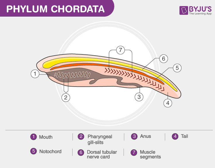
Notochord is a flexible rod-like structure that is found in all chordates. It is similar to cartilage and is involved in signalling and organisation of the nervous system. The notochord usually develops around 16 days after gastrulation from the axial mesodermal cells and is completely formed by the end of the fourth week.
Below is a well-labelled diagram of notochord as found in the Phylum chordata.
Notochord Diagram

Features
- It is a cellular structure that runs along the longitudinal axis of the embryo and ventral to the central nervous system.
- It is composed of glycoproteins that are packed in a sheath of collagen fibres that has been wound into two opposing helices.
- The glycoproteins are deposited in vacuolated and turgid cells.
- The angle between the collagen fibres determines lengthening and thinning of the structure or shortening and thickening of the structure.
- Vertebrates possess the notochord in the embryonic stages but soon develop into an ossified vertebral column with intervertebral discs.
- Any animal that possesses notochord in its life are called chordates and those that do not are called non-chordates.
- The primary function of the notochord is signalling that helps in transformation of unspecified embryonic cells into specific tissues and organs.
- It is also involved in the development of the central nervous system. It signals and activates proteins that induce the formation of motor neurons.
- Apart from this, the notochord gives structural support to the animal’s body. It also helps in locomotion by the contraction of muscle fibres.
Visit BYJU’S Biology for more important information.
Also Read:
Comments