Almost 80% of our sensory responses from the environment is said to be perceived through the eyes. This processing of visual perception holds one-quarter of one’s brain. The process of visual perception is a highly complicated process. It comprises three main sections –
- Optics of the eye
- Detection of photon and the first image processing in the retina
- Signal transmission and hence the processing of the visual cortex of the brain
Structure of the EyeDifferent Parts of the EyePhysiology of VisionFrequently Asked Questions
Structure of the Eye
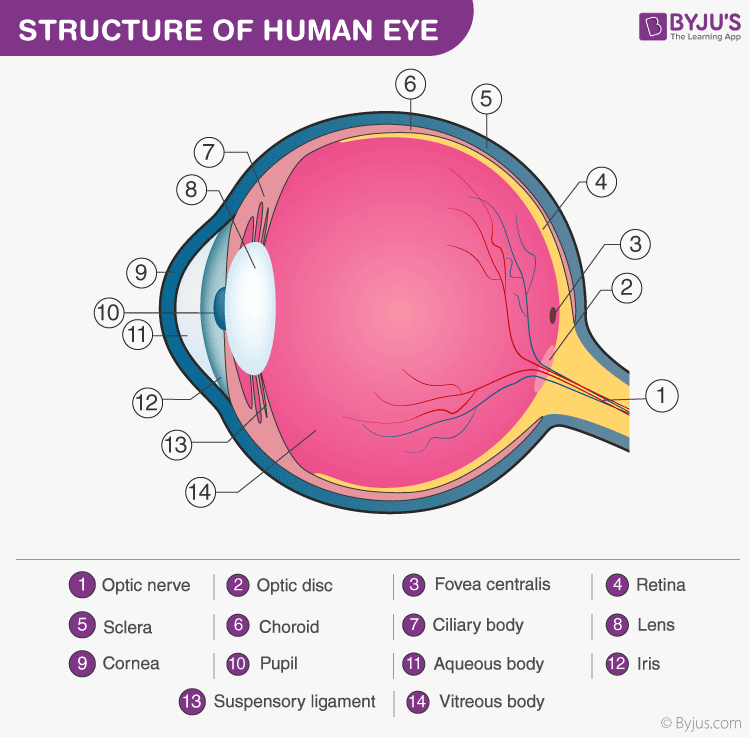
Although the eye is a small structure, it is the most complex organ of the human body. The eye is placed in a bony socket in the skull which extends only a small part outside that is visible. The eye wall comprises three layers – innermost, outermost and the middle layer.
Innermost layer –
Here is the retina, it can be seen located directly behind the eyeball. The capillary found in the middle choroid nourishes the retina when available. It is the light sensitive part of the eye because it has many photoreceptors that are of two kinds (cones and rods). Rods are accountable for white and black visions and functional to see at night. Cones, on the other hand, account for different colour visions.
Middle layer –
It is the choroid comprising black pigmented cells, richly supplied with blood capillaries. This layer forms the ciliary body and iris.
Outermost layer –
It is also referred to as the sclera. It is a whitish tough layer comprising tissues which are connected together. This sclera functions to primarily protect and maintain the shape of the eyeball.

Recommended Video:

Different Parts of the Eye
The eye comprises several structures, take a look at the table for details of each structure.
| Eye structure | Description |
| Cornea | It is a domed shaped structure shielding the eye against anything which can cause harm to the eye. |
| Lens | It is a very transparent layer, post the pupils takes in ambient light, then the lens focuses the light onto the retina. |
| Sclera | It forms the outermost part of the eye. It is white in appearance and is accountable to maintain the eyeball’s shape. |
| Retina | Located at the back of the eye. Its main role is in receiving light from the focus and passing it to electrical impulses before it reaches the brain. |
| Pupil | It is seen at the eye’s center. It is like a black dot having a tiny hole which enables light to pass. |
| Choroid | Forms the interphone between the sclera and the retina accountable for rendering nutrients to other portions of the eye. |
| Macula | Found in proximity to the retina. It aids the eye to focus on an object. |
| Conjunctiva | Conjunctiva gland is the part comprising mucus to moisten the eye. It aids in always keeping the eye moist. In the event of malfunction or failure of this gland, serious itching or pain can occur. It is also responsible to protect the cornea. |
| Iris | Colour of the eye is determined by this. This part imparts the eye with the colour. It surrounds the pupils by all sides. The iris shrinks and widens the pupils based on the light’s intensity entering the eye. The iris widens the pupil if the light is low and vice versa. |
| Optic nerves | Bundle of nerves carrying impulses to the brain from the retina. |
| Anterior and posterior chambers | The front part in the interior of the eye section forms the anterior chamber and the back part forms the posterior chamber. |
Physiology of Vision
Visual process is the series of actions that take place during visual perception. During the visual process, the image of an object seen by the eyes is focused on the retina, resulting in the production of visual perception of that object.
The physiological events which take place are as follows –
- Light’s refraction which enters the eye
- Image focuses on the retina by accommodation of lens
- Image convergence
- Photochemical activity in the retina and the conversion into neural impulse
- To process in the brain and then perception
All the parts of the eye function together thus enabling us to see. At first light enters through the clear front layer of the eye, the cornea. Due to its structure (dome-shaped), it bends light to aid the eye in focusing.
Some part of this light passes the eye through the pupil opening. The coloured part of the eye, the iris, regulates how much light enters the pupil.
Light enters through the lens then when the lens functions with the cornea to focus light aptly on the retina. When light passes the retina, special cells referred to as photoreceptors convert light into electrical signals. These signals pass from the retina to the brain through the optic nerve. The brain then turns signals into images which we see.
Read more: Visual Pathway flowchart
Explore more related concepts important for NEET preparation at BYJU’S.
Frequently Asked Questions
What are the components that make the visual pathway?
The visual pathway consists of optic nerves, retina, optic tracts, optic chiasm, optic radiations, visual cortex and the lateral geniculate bodies.
What is the physiology of sight?
While looking at an object, light rays from that object are refracted and brought to a focus upon the retina. The image falls on the retina in an inverted position and the energy in the visual spectrum is converted into electrical impulses by rods and cones of the retina. Impulses from rods and cones reach the cerebral cortex through the optic nerve and the sensation of vision is produced in the cerebral cortex.
What are the functions of rods and cones?
Rods are very sensitive to light and have a low threshold. So, the rods are responsible for dim light vision or night vision or scotopic vision. Cones have a high threshold for light stimulus. So, the cones are sensitive only to bright light. Therefore, cone cells are called receptors of bright light vision or daylight vision or photopic vision.
Don’t miss:
| Structure of Eye MCQs for NEET |
| Human Brain MCQs for NEET |
Comments