Epithelium Definition
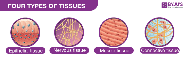
A membranous animal tissue composed of epithelial cells tightly packed together and connected by cell junctions is known as the epithelium. Layers of “epithelium” cells line hollow glands and organs. The outer layer of the body is composed of epithelial cells. A basement membrane lies beneath the layer(s) of epithelial cells.
The internal and external organs of the body are lined and covered in epithelial tissue. Epithelial tissue, for example, lines the interior walls of blood vessels, forms the skin, and covers the liver. Many different forms of epithelial cells comprise the epithelial tissues, including square, flat, round, and columnar.
Transitional epithelial tissue is a particular type of epithelial tissue that has many layers of cells rather than one. The stratified epithelium is an epithelial tissue made up of numerous cell layers. The components of the urinary system are lined with cells of the transitional epithelium.

Table of Contents
- What is Transitional Epithelium
- Transitional Epithelium Structure
- Transitional Epithelium Function
- Transitional Epithelium Examples
- Frequently Asked Questions (FAQs)
What is Transitional Epithelium
Transitional epithelium is a type of stratified epithelium composed of several layers of cells, with the morphology of cells varying depending on the function of the organ. In a relaxed state, the epithelium appears spherical or cubical, except for the apical layer, which may flatten when stretched. Since this epithelium is primarily present in the urinary system, it is sometimes known as “urothelium.”
This epithelium can be found in the ducts of the prostate gland as well as lining the ureters, urinary bladder, and urethra.
Transitional Epithelium Histology
The lower urinary tract comprises transitional epithelium (urothelium), a unique stratified epithelium. It changes from a thicker to a thinner epithelium rapidly to accommodate distention and contraction.
Relaxed (non-stretched)
The surface of the transitional epithelium is covered in a diverse range of cells, including enormous, dome-shaped umbrella cells. They are known as umbrella cells since they protect numerous epithelial cells beneath them. Active umbrella cells are present at the luminal layer.
Extended (stretched)
A thinner transitional epithelium develops and the umbrella cells expand and get flattened.
Also, read: Epithelial Tissue
Transitional Epithelium Structure
The structure of transitional epithelium varies depending on the cell layer. While the superficial layer’s cells have various shapes depending on the degree of distension, the basal layer’s cells are either cuboidal (cube-shaped) or columnar (column-shaped).
- Transitional epithelia are composed of three to four layers of cells, with the lowest or basal layer remaining in connection with the basement layer.
- The basal layer, the lowest layer, supplies the higher layers’ ongoing cell regeneration.
- The cytoplasm of basal layer cells is rich in tonofibril-forming proteins, converging with hemidesmosomes to produce strong contact with the basement layer.
- The basal layer cells have an excess of mitochondria because they require more energy to rebuild the epithelium.
- The middle layer or the intermediate layer, is composed of fast dividing, proliferative cells providing rapid cell renewal in the event of damage or injury to the existing cells.
- There are many cells in this layer with the Golgi apparatus, which helps transport proteins such as keratin to superficial layers.
- High levels of keratinisation in the superficial layer cells act as a barrier against water and salts.
- The apical layer or the superficial layer, covers the lumen and guards the underlying layer of cells against harmful pathogens and waste products entering the lumen.
- Desmosomes and gap junctions are used to link the cells in the epithelium. These structural components permit the epithelium to grow, making the cells more fragile.
- The transitional epithelium is avascular, lacking a blood vessel supply, similar to all other epithelial tissues. The blood arteries of the surrounding connective tissues supply nutrition, oxygen and excrement to the cells of this epithelium.
- The cells do, however, have a specific nerve supply.
Transitional Epithelium Function
The transitional epithelium serves two basic functions, determined by the composition and structure of the cell:
Barrier to Permeability
- The tissue offers high impermeability to water and various substances because of the significant keratin deposits present in the cells.
- Even when fully stretched, the transitional epithelium cells offer resistance to osmotic pressure, which prevents desiccation.
- Additionally, the bloodstream is protected from chemicals and toxins entering.
- The urinary system serves as a typical example of this function, as the urothelium’s cells do not become dehydrated even in the presence of hypertonic urine in the lumen.
Control of Volume
- The second essential function of these cells is the ability to stretch and expand according to fluid pressure.
- Cells in the superficial layer of the excretory system stretch, altering their form from round to flat, as a result of increased fluid volume in the urine bladder and ureters.
- Stretching increases organ volume while protecting underlying tissue from exposure to urine toxins.
Transitional Epithelium Examples
The urothelium is the most common form of transitional epithelium. The transitional epithelium, also known as the urothelium, lines the urethra, ureters, and urinary bladder.
The transitional epithelium that lines the prostatic urethra of the male reproductive system is connected with the urothelium of the urinary bladder.
Related Links:
Main Page: BYJU’S NEET
Comments