Find below the important notes for the chapter, Body Fluids and Circulation, as per NEET Biology syllabus. This is helpful for aspirants of NEET and other exams during last-minute revision. Important notes for NEET Biology- Body Fluids and Circulation covers all the important topics and concepts useful for the exam. Check BYJU’S for the full set of important notes and study material for NEET Biology and solve the NEET Biology MCQs to check your understanding of the subject.
Download Complete Chapter Notes of Body Fluids and Circulation
Download Now

| Name of the NEET sub-section | Topic | Notes helpful for |
| Biology | Body Fluids and Circulation | NEET exams |
Body Fluids and Circulation – Important Points, Summary, Revision, Highlights
Blood ComponentsBlood GroupsCoagulationCirculatory PathwaysHuman Circulatory SystemCardiac CycleECGDouble CirculationRegulationDisorders
Body Fluids and Circulation
Blood

It is a fluid connective tissue composed of different cells (RBCs, WBCs and platelets). The pH of blood is ~7.4, i.e. slightly alkaline.
- Plasma constitutes 55% of blood volume. It is a viscous fluid and contains 90-92% water. Other than water, plasma contains 6-8% proteins (fibrinogens, albumins and globulins), amino acids, glucose and small amounts of electrolytes (Na+, Ca++, Cl–, etc.).
- Formed Elements include erythrocytes, leukocytes and blood platelets
Erythrocytes or RBCs are the most abundant cells present in the blood
>> formed in the bone marrow
>> biconcave and without a nucleus
>> have a lifespan of 120 days and get destroyed in the spleen (RBCs’ graveyard)
Leucocytes or WBCs are colourless due to the absence of haemoglobin. They are of two types granulocytes and agranulocytes.
| Also see: NEET Answer Key 2022 |

>> have a life span of 3-4 days
>> granulocytes include neutrophils, eosinophils and basophils
>> agranulocytes include lymphocytes and monocytes
>> neutrophils have a polymorphic nucleus and have phagocytic ability
>> basophils are involved in inflammatory reactions as they secrete histamine, serotonin
>> eosinophils are involved in an allergic reaction
Blood platelets or thrombocytes are involved in the clotting of blood. They are formed from the megakaryocyte cells of bone marrow.
Blood Groups

ABO grouping: There are four types of blood group, A, B, AB and O. It is based on the presence or absence of surface antigens on RBCs (antigen A and B).
| Blood Group | A | B | AB | O |
| Antigen on RBCs | A | B | A, B | nil |
| Antibodies in plasma | Anti-B | Anti-A | nil | Anti-A, Anti-B |
| Donor blood type | A, O | B, O | A, B, AB, O
(universal acceptor) |
O (universal donor) |
Rh grouping
80% of people have Rh antigen on the surface of RBC. Those who have Rh antigen are called Rh+ and those without it are Rh–.
Erythroblastosis fetalis: When the mother is Rh– and the father is Rh+ and the foetus blood group is Rh+, then there are chances of mixing of foetus’ blood with that of the mother’s blood during delivery. The mother’s body produces antibodies against the antigen, which may go to the foetus and result in the agglutination of RBCs in the subsequent pregnancy. To avoid this mother is given anti-Rh antibodies after the delivery of the first child.
Coagulation of Blood
Blood clotting prevents excess loss of blood from an injury. The clot is made up of a network of fibrins, which traps dead formed elements.
Inactive fibrinogen → Fibrin by the enzyme Thrombin
Inactive prothrombin → Thrombin by the enzyme Thrombokinase
An injury stimulates platelets to release certain factors, which initiate the blood coagulation process.
Ca++ plays an important role in the process of coagulation.
Lymph
Lymph is also a fluid connective tissue. The lymphatic system drains back the interstitial fluid back to major veins. Lymph consists of lymphocyte cells and is part of the immune system of the body. Fats are absorbed in the intestine by lacteals of villi and transported to the blood by lymph.
Circulatory Pathways
There are two types of circulatory systems:
- Open circulatory system: Blood vessels are not found and blood is present in the open cavity known as sinuses, where internal organs float. This type of system is present in arthropods and molluscs.
- Closed circulatory system: Blood vessels circulate the blood. This type of system is present in Annelids and Chordates.
| Name | Type of Heart | Circulation |
| Fish | 2 chambered, 1-atrium and 1-ventricle | Single circulation |
| Amphibian and Reptiles | 3 chambered, 2-atrium and 1-ventricle | Incomplete double circulation |
| Crocodiles, Birds and Mammals | 4 chambered, 2-atrium and 2-ventricle | Double circulation |
Human Circulatory System
Heart- Four chambered, derived from mesoderm. It is present between the two lungs in the thoracic cavity and protected by the pericardium.
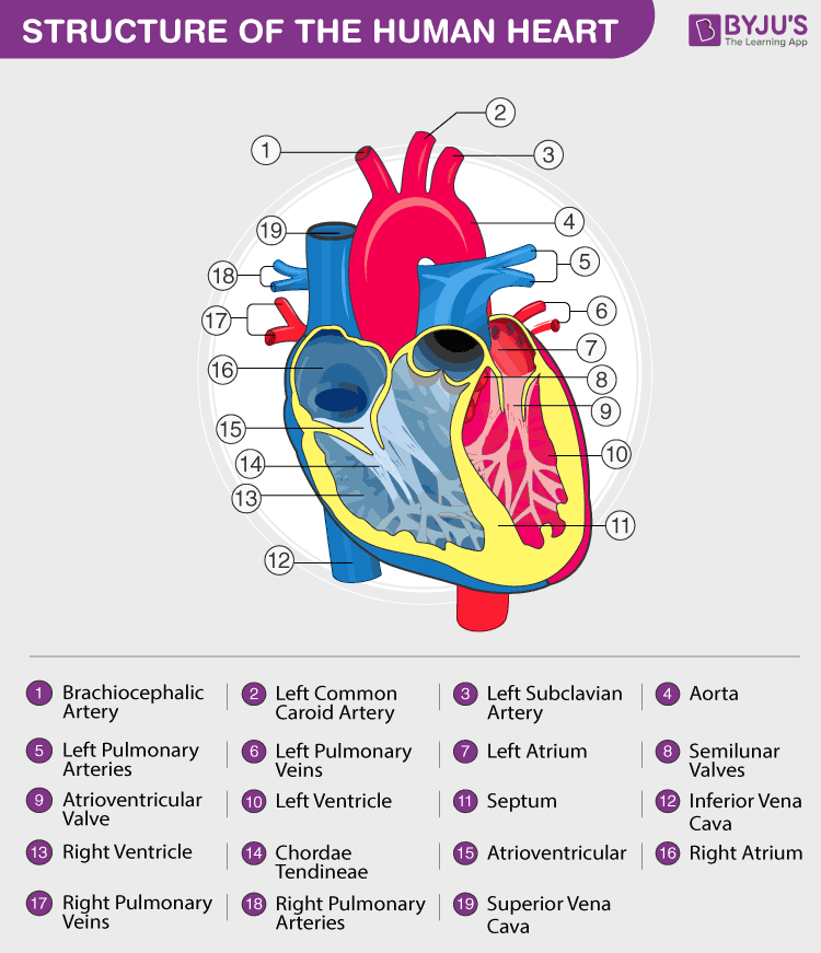
- A bicuspid (mitral) valve is present between the left, atrium and ventricle
- A tricuspid valve is present between the right, atrium and ventricle
- The right ventricle opens into the pulmonary artery and the left ventricle opens into the aorta. Both the openings are guarded by semilunar valves
- The heart is made up of cardiac muscles. There are nodal tissues also present in the heart.
- The sinoatrial node (SAN) is present on the upper right corner of the right atrium and the atrioventricular node (AVN) is present at the lower-left corner of the right atrium
- The AVN divides into right and left bundle at the interventricular septum and branches into minute fibres, that are present throughout the ventricle and are called Purkinje fibres
- The nodal musculature is auto-excitable, i.e. it can generate action potential without any external stimuli
- Bundle of His- Purkinje fibres along with the AV bundles form the bundle of His. It regulates the heartbeat and conducts the electrical impulses
- The SAN is called ‘pacemaker’, it initiates and maintains the heart’s rhythmic activity. It can generate 70-75 action potentials per minute
- Arteries and veins are together called blood vessels, which transport blood to various parts of the body and bring back to the heart
- Arteries and veins are made up of three layers
- Inner tunica intima – squamous endothelium
- Middle tunica media – elastic fibres and smooth muscles (thin in the veins)
- Outer tunica externa – fibrous connective tissue having collagen fibres
Cardiac Cycle

- At the start of the cycle, all the four chambers remain in a relaxed state known as joint diastole
- Joint diastole is followed by atrial systole, on the generation of action potential from SAN node
- The action potential then gets transferred to AVN and then to the bundle of His leading to the contraction of the ventricle, i.e. ventricular systole and at the same time atrial diastole occurs
- Ventricular systole causes closure of tricuspid and bicuspid valves. The semilunar valves open resulting in the flow of blood to vessels in the circulatory pathways
- Ventricular diastole follows with the closure of semilunar valves
- Then bicuspid and tricuspid valves open and the state of joint diastole is achieved once again
- This completes the one full cycle of the cardiac cycle
- The SAN generates a new action potential and the full process repeats
- As the heart beats ~72 times per minute, so there are ~72 cardiac cycles in a minute
- Stroke Volume is the amount of blood each ventricle pumps in a cardiac cycle, i.e. 70 ml
- Cardiac Output is the total output of blood from each ventricle in a minute, i.e. stroke volume multiplied by the no. of heartbeats per minute, which is ~5 L in a healthy individual. The cardiac output varies from person to person. An athlete will have much more cardiac output compared to an ordinary person
- In each cardiac cycle, two distinct sounds are produced, ie. ‘lub’ and ‘dub’, which can be heard using a stethoscope
- The first sound ‘lub’ is produced when bicuspid and tricuspid valves close at the time of ventricular systole
- The second sound ‘dub’ is produced when semilunar valves close at the time of ventricular diastole
ECG

The electrical activity of the cardiac cycle can be recorded in a graphical form by electrocardiogram or ECG.
Various phases of each cardiac cycle are represented by a letter from P to T.
- P wave → excitation or depolarisation of atria
- QRS complex → depolarisation of ventricles
- T → repolarisation of ventricles (excited to normal)
The no. of QRS complex recorded in a time period tells the heart rate of the patient. Any abnormality or disease can be diagnosed by ECG if the graph shows any deviation from a regular pattern.
Also Read: MCQs on ECG
Double Circulation
Pulmonary Circulation: The pulmonary artery receives deoxygenated blood and transports it to the lungs. From the lungs, oxygenated blood is transported to the left atrium via the pulmonary vein. This pathway is called the pulmonary circulation.
Pulmonary artery – deoxygenated blood, Pulmonary vein – oxygenated blood.
Systemic Circulation: The oxygenated blood goes to the aorta from the left ventricle. The oxygenated blood then gets transported from the aorta to various tissues by arteries, arterioles and capillaries.
The deoxygenated blood from various parts of the body comes to the right atrium by the network of venules, veins and vena cava.
This pathway is called systemic circulation.
Hepatic Portal System: The vascular connection between the liver and digestive tract. The hepatic portal vein collects intestinal blood and transports to the liver and then it goes to the systemic circulation.
Regulation of Cardiac Activity
The heart is myogenic, as its activity is regulated by nodal tissues.
The heart’s function is moderated through ANS (autonomic nervous system) by a neural centre in the medulla oblongata.
Sympathetic neural signals and adrenal medullary hormones – increase heart rate and cardiac output.
Parasympathetic neural signals – decrease heart rate
Disorders of the Circulatory System
High Blood Pressure (Hypertension): The normal blood pressure of a human being is 120/80 mm Hg. 120 is the systolic pressure and 80 is the diastolic pressure. High bp > 140/90 mm Hg for a longer duration may lead to various heart diseases and may also adversely affect the brain and kidneys.
Coronary Artery Disease (CAD): It is also referred to as atherosclerosis. It affects the supply of blood to the heart. The lumen of the arteries become narrower due to deposition of cholesterol, fat, calcium or fibrous tissues.
Angina (Angina Pectoris): The severe chest pain due to the scarcity of oxygen reaching the heart muscles, when there is interrupted blood flow.
Heart Failure: When the heart fails to pump enough blood to meet the requirement of the body. Lungs congestion is one of the symptoms of the disease. It is different from a heart attack, where there is sudden damage of heart muscle due to an insufficient blood supply or cardiac arrest when the heart stops beating.
Coronary Thrombosis: It is a condition when there is a clot formation in the coronary artery. It happens frequently in the left anterior descending coronary artery.
Explore the next chapter for important points with regards to the NEET exam only at BYJU’S. Check the NEET Study Material for all the important concepts and related topics.
Also Check:
NEET Flashcards: Body Fluids And Circulation
NEET Flashcards: Excretory Products And Their Elimination
NEET Flashcards: Locomotion And Movement
NEET Flashcards: Neural Control And Coordination
NEET Flashcards: Chemical Coordination And Integration
Recommended Videos:
Session-1

Session-2
Session-3

Session-4
Also see:
Comments