


1
Question
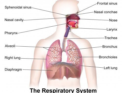
The figure shows a diagrammatic view of the human respiratory system. Label its parts.

The figure shows a diagrammatic view of the human respiratory system. Label its parts.
Open in App
Solution
A: Trachea
- The trachea rises below the larynx and moves down the neck.
- The walls of the trachea are made up of C-shaped cartilaginous rings providing hardness to the trachea and maintaining it by helping it completely expand.
- The trachea extends further down splitting into two bronchi, one for each lung.
B: Pleural membrane
- A pleura is a serous membrane with two layers of membranous tissue that folds back on itself to produce a pleural sac.
- The parietal pleura, which connects to the chest wall, is the name of the outer layer.
- The lungs, blood arteries, nerves, and bronchi are all covered by the inner layer, which is known as the visceral pleura.
C: Alveoli
- The bronchioles get terminated into balloon-like structures, the alveoli.
- The alveoli are single-celled sacs of air with thin walls.
- It facilitates the exchange of oxygen and carbon dioxide molecules into or away from the bloodstream.
Other parts of the human respiratory system:
Lungs:
- These are the primary organs of respiration in humans as well as other vertebrates.
- They are present in the thoracic cavity of the chest.
- The primary function of the lungs is to carry out the exchange of gases between the blood and the air.
Nasal cavity:
- This is lined with hair and mucus to filter the air inhaled from dust and dirt.
Larynx:
- Two cartilaginous chords lay to make the framework of the larynx.
- It is present in front of the neck and is responsible for vocals as well as helps in respiration, therefore also referred to as a voice box.
- When food is swallowed, epiglottis folds over the top of the windpipe thereby preventing food from entering the larynx.
Bronchi:
- The trachea splits into two tubes called bronchi.
- The bronchi divide into secondary and tertiary bronchioles and branch out into small air sacs called the alveoli.
Suggest Corrections


1


Similar questions


Join BYJU'S Learning Program


Explore more
Join BYJU'S Learning Program


