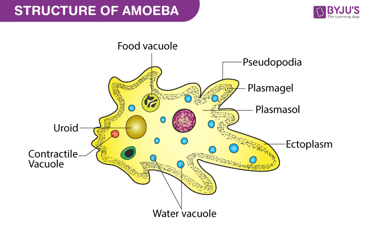
Pseudopodia (sing: pseudopodium) are cytoplasmic projections that are found on the cell membranes of eukaryotic cells. The word pseudopodia is derived from pseudo meaning false and podia meaning feet.
Below is a well-labelled diagram of an amoeba which shows the cytoplasmic projections of pseudopodia. Scroll down for more on pseudopodia.

Features
- The arm-like temporary projections arising from the eukaryotic cell membrane in the direction of movement are referred to as pseudopodia.
- The pseudopodia are made up of actin filaments, microtubules and intermediate filaments. They are filled with cytoplasm.
- Pseudopodia are commonly found in amoebas. They can have either a single pseudopodium present (example: Entamoeba histolytica) or numerous projections arising from the cell membrane (example: Amoeba proteus).
- Pseudopodia are also formed by cells of higher animals such as white blood cells.
- The main function of pseudopodia is to help in locomotion and feeding. They also help in sensing targets which can then be engulfed, such a type of pseudopodia are called phagocytosis pseudopodia.
- Pseudopodia gives amoeboid movement to the cells which is a sliding or crawling-like form of locomotion.
- During feeding, the extensions either surround the prey and engulf it or trap it on a sticky mesh like structure.
Types of Pseudopodia
Based on appearance, there are four types of pseudopodia –
- Filopodia
- Lobopodia
- Reticulopodia
- Axopodia
Filopodia are slender branching structures that have a clear and translucent appearance. The branching structure shows contraction of the microfilaments which makes the sliding movement of the cells against a surface very easy, even if it is relatively heavy.
Lobopodia are flattened and cylindrical bulbous structures that are commonly found in amoebas. These pseudopodia contain both endoplasm and ectoplasm layers. The movement in this type of pseudopodia is advanced by the contraction of ectoplasm which leads to the forward flow of endoplasm and the whole body moves forward.
Reticulopodia are formed by fine threads that anastomose to form a dense network. This dense network of filaments helps in easy engulfing and trapping of the prey.
Axopodia are the most complex type of pseudopodia found. They are made up of an outer layer of flowing cytoplasm with bundles of microtubules present at the core that are cross linked in a specific manner. These structures phagocytose the prey by retracting as soon as they sense a physical contact.
Visit BYJU’S Biology for more information.
Also Read:
Comments