NEET 2023 is approaching, and it is time to do thorough revisions and give your best for the entrance exam. There are often some important tables and charts for NEET Biology which we tend to overlook while reading the theory. Here we have combined all the important tables and charts for your revision, chapterwise.
Class 11
Chapter 2 – Biological Classification
Table 1
| Kingdoms | Monera | Protista | Fungi |
| Cell Type | Prokaryotes | Eukaryotes | Eukaryotes |
| Cell Wall | Noncellulosic
(Polysaccharide + amino acid) |
Present in some (varied compositions) | Present with chitin |
| Nuclear Membrane | Absent | Present | Present |
| Body Organisation | Cellular | Cellular | Multicellular / Loose tissue |
| Mode of Nutrition | Autotrophic (Chemosynthetic and Photosynthetic) and Heterotrophic (parasitic / saprophytic) | Autotrophic (Photosynthetic) and Heterotrophic | Heterotrophic (Saprophytic / Parasitic) |
R.H Whittaker gave the five kingdoms of classification and made morphological characters the basis for this division. This table raises multiple MCQs based on the kingdoms’ cell wall, cell type and mode of nutrition. Let us revise them one by one.
Cell Type: The Monera kingdom that consists of bacteria is considered a prokaryote because it has a primitive nucleus and no nuclear membrane. Protista and Fungi, on the other hand, are both eukaryotes because they possess a distinct nuclear membrane.
Cell Wall: Monera has a non-cellulosic cell wall made up of NAG and NAM peptidoglycans. Protists may or may not have a cell. If they possess a cell wall it is mostly cellulosic but other components vary among different species. All the fungal cell walls are made up of chitin.
Body Organisation: All the bacterial species found in the Monera kingdom are unicellular cells. One striking difference between Protista and Fungi is that both are eukaryotes (eu meaning true nucleus) but Protists are unicellular and Fungi are multicellular.
Mode of Nutrition: Bacteria, resident of the lowest kingdom Monera, possess the widest range of nutrition, meaning it can feed on anything and everything. It can be autotrophic bacteria where it can make its own food (photosynthetic) or it can create energy by the oxidation of inorganic compounds. It can be heterotrophic as well, where it acts as either a parasite or a saprotroph. Protists are mostly photosynthetic (e.g., diatoms and dinoflagellates) but some are heterotrophic as well. Last, the Fungi are strictly heterotrophic, they never make their own food. They either extract nutrition from other organisms (parasitic) or feed on dead material (saprophytic)
Table 2
| Kingdoms | Plantae | Animalia |
| Cell type | Eukaryotic | Eukaryotic |
| Cell wall | Present (Cellulose) | Absent |
| Nuclear
membrane |
Present | Present |
| Body
organisation |
Tissue / organ | Tissue / organ / organ system |
| Mode of
nutrition |
Autotrophic
(Photosynthetic) |
Heterotrophic
(Holozoic/Saprophytic etc.) |
The other two kingdoms of the five kingdom classification are Plantae and Animalia. They are the most advanced kingdoms and are a source of some very important questions.
Striking Differences between Plantae and Animalia
- The plants have a cellulosic cell wall, whereas animals do not possess any cell wall.
- The body organisation in plants is limited to tissues and organs, whereas in animals, they have tissues, organs and a well-designed organ system.
- Plants are photosynthetic organisms that make food for the entire planet. They have an autotrophic mode of nutrition. Animals, on the other hand, are heterotrophic organisms that depend on plants and other animals for their food.
Chapter 3 – Plant Kingdom
Table 3
| Rhodophyceae | Phaeophyceae | Chlorophyceae | |
| Common Name | Red algae | Brown algae | Green algae |
| Major Pigments | Chlorophyll a, Chlorophyll d. Phycoerythrin | Chlorophyll a, Chlorophyll c,
Fucoxanthin |
Chlorophyll a, Chlorophyll b |
| Stored Food | Floridean Starch | Laminarin, Mannitol | Starch |
| Cell Wall | Cellulose, pectin and polysulphate esters | Cellulose and algin | Cellulose |
| Flagellar number and position of insertions | Absent | 2, unequal, lateral | 2-8, equal, apical |
| Habitat | Fresh water, brackish water, saltwater | Fresh water (rare), brackish water, salt water | Fresh water, brackish water, saltwater |
Algae are chlorophyll-bearing aquatic photosynthetic organisms. They occur in a wide variety, size, form and range. Based on the types of pigments present, the algae can be divided into Rhodophyceae, Phaeophyceae and Chlorophyceae. However, the basis of differentiation between the three types of algae is not limited to their pigment types, they differ in various other morphological characteristics. Let us revise them.
Pigments Present: The pigment chlorophyll a is found in all the three types of algae. Red algae or rhodophyceae have chlorophyll d and phycoerythrin which imparts red colour to the organism. Similarly, phaeophyceae or brown algae have chlorophyll c and fucoxanthin that imparts the brown colour to the algae. Chlorophyceae are very similar to higher plants as they contain both chlorophyll a and b which are the same as found in higher plants.
Stored Food: Rhodophyceae stores its food in the form of floridean starch. Phaeophycaeae stores food in the form of laminarin and mannitol that are complex carbohydrates. Chlorophyceae, on the other hand, have storage bodies called pyrenoids that store food in the form of starch, just like higher plants.
Cell Wall: Rhodophycaeae have cell walls made up of cellulose, pectin and polysulphate esters. The polysulphate esters are used to extract agar-agar, which is used commercially. The red algae also yield hydrocolloids such as carrageen that are water-holding substances. Phaephycaeae have cell walls made up of cellulose and algin. Algin is extracted and used commercially, which is another hydrocolloid. Chlorophyceae, again, is similar to higher plants and has a cell wall made up of cellulose.
Flagellar number and position of insertions: Rhodophyceae is a non-motile algae that has no flagella present on its body. They remain attached to the substratum. Phaeophyceae have two unequal flagella that arise laterally (sideways) from their bodies. Chlorophyceae have 2-8 equal and apically arising flagella.
Habitat: Chlorophyceae are found in both freshwater and seawater. Phaeophyceae and Rhodophyceae, on the other hand, are mostly marine.
Table 4
| Life Cycle | Haploid:Diploid |
| Haplontic | Haploid gametophyte stage is predominant. E.g, Algae |
| Diplontic | Diploid sporophytic stage is predominant. E.g, Angiosperms and Gymnosperms |
| Haplo-diplontic | Either haploid gametophyte or diploid sporophyte is slightly dominant. E.g, Bryophytes and Pteridophytes, respectively. |
Plants have different stages in their life cycle which is known as alternation of generation. It produces two different types of bodies – haploid and diploid. The haploid gametophyte, as the name suggests, produces gametes by mitosis. The gametes fuse to form a diploid zygote that again divides by mitosis to give rise to diploid sporophyte. The diploid sporophyte then divides by meiosis to give rise to haploid spores that germinate into haploid gametophytes. In this way, plants alternate between the haploid and diploid stages and show either haploid, diploid or haplodiplontic phases of life cycle.
Haplontic Life Cycle: Algae show haplontic life cycle where haploid gametophyte stage is predominant. The sporophyte does not exist freely. It is seen as a diploid one-celled zygote that eventually divides by meiosis to form haploid spores that germinate into haploid gametophytes. Fucus is an algae that shows diplontic life cycle.
Diplontic Life Cycle: Higher seed-bearing plants like angiosperms and gymnosperms show a diplontic life cycle. In this type, diploid sporophytic phase is the predominant and independent phase of life. Gametophytic generation is short-lived and dependent on sporophyte.
Haplo-diplontic Life Cycle: In this type, either the sporophyte or gametophyte is a slightly dominant phase of their life cycle. In bryophytes, an independent, erect and photosynthetic haploid gametophyte is the dominant stage or the parent stage. The sporophyte is short lived and dependent on the gametophyte for its anchorage and nutrition.
Conversely, in Pteridophytes, the diploid, independent and photosynthetic sporophytic body is the dominant phase or the parent stage of the life cycle. The gametophyte is independent, but short-lived.
Ectocarpus, polysiphonia and kelps are some algae that show a haplo-diplontic life cycle.
Chapter 5 – Morphology of Flowering Plants
Root Modifications
Table 5
| Tap Root | Adventitious Root |
| Conical Root (carrot) | Tuberous Root (sweet potato) |
| Fusiform Root (radish) | Prop Roots (Banyan tree) |
| Napiform Root (turnip) | Stilt Roots (Maize, Sugarcane) |
| Tuberous Root (4 o’clock plant) | |
| Pneumatophores (Rhizophora) |
Roots are plant organs that provide anchorage and nutrients to the plants. The direct elongation of the radicle leads to formation of primary roots which bear several secondary roots. The primary roots and its branches are referred to as taproots found in dicots. Roots that do not arise from the radicle but any other parts of the plants are referred to as adventitious roots, found mostly in monocots. These roots change their shapes and get modified for storage, respiration and supportive purposes. Let us look at the examples of such root modification that are important for NEET Biology 2023.
Tap roots are modified into different shapes such as conical root (carrot), fusiform root (radish), napiform root (turnip) and tuberous root (4 o’clock plant). These roots are modified for storage purposes and are also consumed by humans as a food source. Another special modification is the formation of pneumatophores or breathing roots in Mangrove plants (Rhizophora). Mangrove plants tend to grow in marshy soil where roots cannot breathe. As a result, the roots become negatively geotropic and arise from the soil to form breathing roots.
Similarly, adventitious roots form tuberous roots in sweet potato, orchids and asparagus for storage purposes. The adventitious roots also modify to provide structural support to monocotyledons. For example – it forms prop roots in banyan trees where roots hang from the aerial branches of the plant to give structural support to the trunk. Stilt roots are formed in maize and sugarcane where supportive roots arise from the lower nodes of the stem.
Table 6
| Underground | Sub-aerial | Aerial |
|
|
|
Similar to roots, stems also modify for different purposes. Stem modifications are very important for NEET Biology because they are also important for vegegtative propagation which you will study in class 12th.
Underground Stem Modifications with Examples: Underground stems modify for food storage purposes that are mainly used by humans for eating. For example – Ginger (rhizome), Potato (Tuber), Colocasia (Corm) and Onion (Bulb).
| Note : Potato is tuberous stem modification whereas sweet potato is a tuberous root modification. |
Sub Aerial Stem Modifications: Sub aerial stem modifications are partially below and partially above the ground. Examples of such modifications include – mint (sucker), Eicchornia (offset), strawberry (stolon) and lawn grass (runner).
Aerial Stem Modifications: Tendrils are aerial stem modifications that are formed from axillary buds. They are slender and coil spirally to help the plant climb. Example – cucumber, watermelon and pumpkin). Thorns are modifications of axillary buds of stems that develop into a straight, pointed and woody structure to protect the plants from animals. Examples – Citrus, Bougainvillaea. Lastly, phylloclade is an important modification that shows both leaf and stem modification. Leaf gets modified into the spine and thus the stem takes over the function of photosynthesis and storage and becomes swollen. Example – Cactus, Opuntia.
Table 7
| Placentation Types | Examples |
| Axile Placentation | China rose, tomato and lemon |
| Parietal Placentation | Mustard, Argemone |
| Free Central Placentation | Dianthus, Primrose |
| Basal Placentation | Sunflower, marigold |
| Marginal Placentation | Pea |
Placentation is the arrangement of ovules within the ovary. There are five types of placentation known – axile, parietal, free central, basal and marginal placentation. Examples of all the five types are important for NEET 2023.

In axile placentation, the ovules are attached in a multilocular ovary and the placenta is present axially. In parietal placentation, the ovules either develop on the periphery of the ovary or inner walls of the ovary. In free central placentation type the ovules are borne on the central axis and there is no septa in the ovary. In basal placentation, the placenta develops at the base of the ovary and only a single ovule is attached to it. Lastly, marginal placentation is characterised by the formation of a ridge by the placenta on the margins of which the ovules are borne.
Chapter 6 – Anatomy of Flowering Plants
Table 8

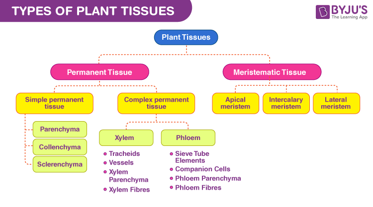
The plant tissues are divided into meristematic and permanent tissues based on their ability to divide or not. These types of tissues are a source of a variety of questions in NEET 2023. Let us revise the important points.
The meristematic tissues are specialised cells that are found in regions of active cell division. They are divided into apical meristem, intercalary meristem and lateral meristem based on the positions they are found in. The apical meristem is found on shoot and root apex. The intercalary meristem is found in the nodal regions of the plant. And lastly, the lateral meristems are found laterally or on the sides of the plant organ.
| Note: On the basis of origin, apical and intercalary meristems are primary meristems whereas lateral meristem is a secondary meristem. |
The permanent tissues are formed when the primary and secondary meristems lose their ability to divide and become structurally and functionally specialised. The permanent tissues are further divided into simple tissues and complex tissues.
The simple tissues are homogenous tissues such as parenchyma, collenchyma and sclerenchyma. Sclerenchyma is a dead tissue.
Complex tissues are heterogeneous tissues that are made up of different cell types. Xylem and phloem, together known as the conducting tissues, are complex tissues that are made up of different cell types.
Xylem is composed of tracheids, vessels, xylem parenchyma and xylem fibres. The only living component of the four is xylem parenchyma. Phloem is composed of sieve tube elements, companion cells, phloem parenchyma and phloem fibres. Out of the four, phloem fibres, also called bast fibres, are dead tissues.
Class 12
Chapter 2 – Sexual Reproduction in Flowering Plants
Table 9

Pollination is the transfer of pollen grains to the stigma of a pistil. The pollen can either be pollinated in the same flower, in a different flower in the same tree or in a different flower in a different tree. Based on the source of pollen, there are two types of pollination – self pollination and cross pollination.
Self pollination is again of two types – Autogamy and Geitonogamy. Autogamy is the transfer of pollen to the stigma in the same flower. Geitonogamy, on the other hand, is the transfer of pollen from one flower to the stigma of another flower in the same tree. Functionally, geitonogamy is cross pollination but since no genetic changes take place, it is placed under self pollination.
Xenogamy is a type of cross pollination where pollen from the flower of one tree is transferred to the stigma of the flower of another tree. This kind of pollination leads to genetic recombination in the offspring.
| Note: Geitonogamy and xenogamy are together referred to as allogamy. |
Chapter 13 – Organisms and Populations
Table 10
| Species A | Species B | Name of Interaction |
| + | + | Mutualism |
| – | – | Competition |
| + | – | Predation |
| + | – | Parasitism |
| + | 0 | Commensalism |
| – | 0 | Ammensalism |
A habitat becomes functional when different species interact with each other for food, shelter and other resources. These population interactions can be of different types and may be negative or positive in nature. Let us look at the types of population interactions with examples that are important for NEET Biology 2023.
- Mutualism: Mutualism is a positive interaction where both the species gain from each other and hence the + signs for the species in the table. Examples – Lichen is a mutualistic relationship between fungus and photosynthesising algae where algae depends on fungus for shelter and fungus depends on algae for food and nutrition. Similarly mycorrhizae is mutualism between fungus and roots of higher plants.
- Competition: Competition is a negative interaction where both the species fight each other (hence the – signs in both species) for resources such as food and shelter. Example – in the South American lakes, the visiting flamingoes and the resident fish compete against each other for their common food, the zooplanktons.
- Predation: Predation is the interaction where one organism kills and preys on other organisms. Hence, the species A which eats on the other is + and the species B which is being eaten is -. Predation is often thought of in a negative sense as to when a tiger preys on a deer but an animal grazing on a tree is also predation.
- Parasitism: Parasitism is an interaction where one species takes benefits from the other species, sometimes without killing the other species. Ticks, fleas and lices are ectoparasites that reside on external surfaces and gain nutrition from the host. Plasmodium is an endoparasite that resides in the gut of the host, extracts nutrition and causes malaria.
- Commensalism: This is a type of interaction where one species benefits from the other while the other species remains neutral or unaffected. Examples – orchids growing as an epiphyte on a mango branch, barnacles growing on the back of a whale, sea anemone and the clown fishes living inside them.
- Ammensalism: Ammensalism is an interaction where one species is negatively affected whereas the other remains neutral or unaffected.
That’s all about chapterwise biology tables and charts that are important for the preparations of NEET 2023. Visit BYJU’S for more important updates on NEET.
Recommended Video:

Recommended Reads:
Comments