According to the CBSE Syllabus 2023-24, this chapter has been renumbered as Chapter 18.
The process through which two or more organs interact and complement the functions of one another is termed coordination. The neural system and the endocrine system coordinate and integrates all activities of the body so that they function in a synchronized fashion. While the neural system provides an organized network of point-to-point connections for rapid coordination, the endocrine system renders a chemical integration through hormones.
Let us learn in detail the process of neural control and coordination.
Neural System
- It comprises highly specialized cells known as neurons which can receive and transmit different kinds of signals.
- In lower invertebrates such as Hydra, a network of neurons is found
- The Neural system in insects consists of numerous ganglia and neural tissues. It is highly developed in vertebrates
Also Read: Human Nervous System
For more information on Nerve Impulse Mechanism, watch the below video

Human Nervous System
The human nervous system is divided into the following parts:
Central Nervous System
- In vertebrates, the central nervous system is hollow, dorsal, and non-ganglionated, whereas, in invertebrates, it is ventral, solid and contains ganglions.
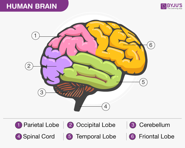
- The central nervous system is further divided into the spinal cord and brain.
Peripheral Nervous System
- It is made up of nerves which extend between the central nervous system and body parts.
- It comprises cranial and spinal nerves.
- It controls all the voluntary functions of the body.
Autonomic Nervous System
- It is made up of nerve fibres and controls the involuntary functions of the body.
- It is composed of the sympathetic and parasympathetic nervous system.
Neurons
- Neurons are composed of three major parts – cell body, dendrites and axon. The cytoplasm of the cell body contains cell organelles and a few granules known as Nissl’s granules.
- Dendrites are the short fibres which repeatedly branch and emerge out of the cell body. They transmit impulses towards the cell body.
- Axons are long fibres whose distal end is branched. Each branch terminated as a bulb-like structure known as a synaptic knob comprising the synaptic vesicles containing neurotransmitters.
- Axons transmit nerve impulses from the cell body to synapse. Neurons are divided into three types depending upon the number of axons- multipolar and two or more dendrites. The axons can be myelinated and non-myelinated.
- The Schwann cells enclose the myelinated nerve fibres and form the myelin sheath around the axon.
- Gaps between two adjacent myelin sheaths are known as nodes of Ranvier.
Conduction of Nerve Impulse
- When neurons are in their resting phase (not conducting any impulse), the axon membrane is more permeable to potassium ions but impermeable to sodium ions and the negatively charged proteins found in the axoplasm.
- Plasma in axons contains a low concentration of sodium ions and a greater concentration of potassium ions and proteins. However, the liquid outside the axon contains a high sodium ion concentration and a low potassium ion concentration, thus forming a concentration gradient.
- Active transport of ions takes place across the membrane by the sodium-potassium pump where three ions of sodium are transported outwards and two ions of potassium move into the cell, as a result of which the outer surface of the membrane turns positively charged while the inner surface gets a negative charge hence the cell is in a polarized state developing a resting potential.
- When a stimulus is applied at the site on a polarised membrane, the membrane becomes freely permeable to sodium ions; hence sodium ions move into the cell. The outer side of the membrane gets negatively charged, while the inner side is positively charged. Now, the membrane is in a depolarized state.
- The electrical potential difference produced across the plasma membrane at this site is known as the action potential.
- This area becomes a stimulus for the neighbouring area of the membrane, which becomes depolarized. The previous membrane gets repolarized due to the movement of sodium ions outside the cell. This is how impulses are conducted.
Transmission of Nerve Impulse
- Nerve impulses are transmitted from one neuron to another neuron through synapses which are formed by membranes of a pre-synaptic and post-synaptic neuron.
- Synapses are of two types – electrical synapses and chemical synapses
- When an impulse reaches the axon terminal, it triggers the movement of synaptic vesicles towards the membrane. The plasma membrane and the vesicles fuse and release neurotransmitters in the synaptic cleft, which in turn bind to specific receptors found on the post-synaptic membranes

Reflex Action And Reflex Arc
- The process of responding to a peripheral nerve stimulation occurs involuntarily, requiring the involvement of a part of the CNS known as a reflex action.
- The reflex pathway consists of at least one efferent neuron and one afferent neuron.
- The afferent neuron receives signals from sensory organs and transmits them to the Central Nervous System.
- The efferent neuron transmits signals from CNS to the effector organ.

Human Eye
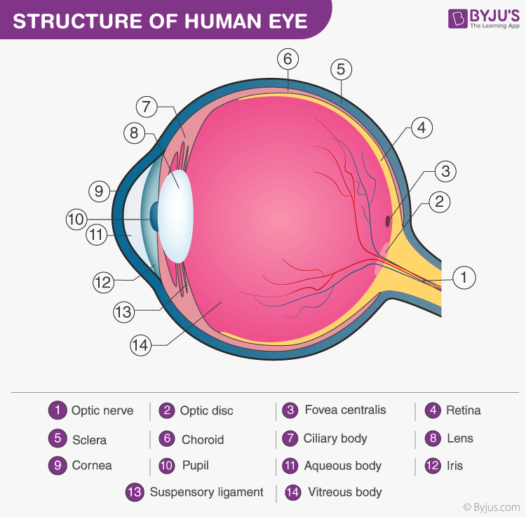
- The eyes are the most important organ of the human body. The wall of the eyeball is composed of three layers – the external layer comprises a dense connective tissue known as the sclera, the anterior portion of it is the cornea and the middle layer choroid, consists of several blood vessels. The ciliary body itself continues forward, forming a pigmented and opaque structure called the iris
- The eyeball consists of a transparent lens held in place by ligaments joined to the ciliary body. The aperture surrounded by the iris in front of the lens is the pupil, whose diameter is regulated by muscle fibres of the iris.

Human Ear
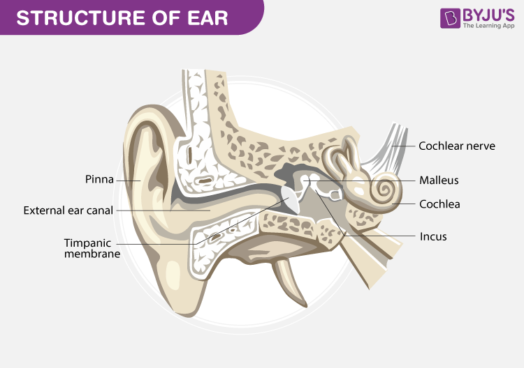
- The ear performs two sensory functions – hearing and maintaining body balance. The human ear can be divided into the outer ear, middle ear and inner ear.
- The outer ear comprises external auditory meatus and pinna. The pinna collects vibrations producing sound, the external auditory meatus extends to the eardrum.

- The middle ear comprises three ossicles called malleus, incus and stapes attached to one another.
- The middle ear cavity is connected with the pharynx through the eustachian tube. It helps in equalising the pressures on either side of the eardrum.
- The inner ear, called the labyrinth, is made up of two parts, the bony and the membranous labyrinths.
Also Read: Control and Corodination
Stay tuned with BYJU’S to learn more through neural control and coordination notes.
Chapter 21 Neural Control And Coordination Notes:- Download PDF Here
Further Reading:
Revision Notes For For Class 11 Biology Chapter 21 Neural Control And Coordination
Important questions for control and coordination
Frequently Asked Questions on CBSE Class 11 Biology Notes Chapter 21 Neural Control and Coordination
What is the function of the Central nervous system?
The central nervous system (CNS) controls most functions of the body and mind. It consists of two parts: the brain and the spinal cord. The brain is the centre of our thoughts, the interpreter of our external environment, and the origin of control over body movement.
What are some facts about the human brain?
1. 60% of the human brain is composed of fat 2. The brain contains about 100 billion neurons and 100 trillion connections 3. The texture of the brain is similar to that of firm jelly
How many parts does the human eye have?
The human eye totally consists of 7 parts that work together.
Comments